
~1.jpg&newheight=260&quality=80)


What is an Ultrasound?
An ultrasound is a procedure that uses ultrasonic waves, known as high frequency sound waves, to produce images of the interior of the body and the internal organs. It works mainly in the same way as sonar. These images are often used in obstetrics, but also for biopsies and echocardiograms, producing either a still or moving image.
Sound waves that travel through different objects is necessary in producing an ultrasound image. To target the organ or area of the body being scanned, an ultrasonic transducer emits ultrasonic waves. When the sound waves hit different tissues, an echo is produced. The transducer then detects the echo and feeds the information into a computer, which then transforms the sound into images. A gel is applied to the skin so the transducer can easily move over it, and allows the transducer to be as close as possible. If the area is tender due to inflammation, a slight pressure may be felt.
After the ultrasound, there are no activity limitations. The images are then interpreted by a radiologist or trained professional who may then give the patient the results. But, often the results are passed on to the patient?s general practitioner. Since radiation is not used, an ultrasound does not have any side effects. Because ultrasound does not easily distinguish between bone and air, it is not used for imaging bones or the lungs.
An ultrasound is commonly used to produce a picture of a baby in the uterus, but can also be used to examine tumors, swelling, cysts, and the heart. The special type of ultrasound that investigates the heart is called an echocardiogram.
What are Ultrasound Tables?
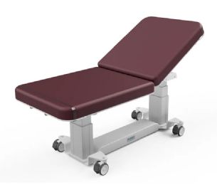
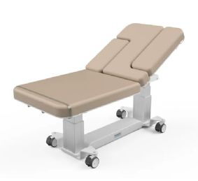
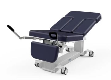
Ultrasound tables are used for various ultrasound exams. They should be adjustable to accommodate multiple users, comfortable for patients, easy to move around with accessible brakes for the sonographer, and designed to be used by a sonographer in the seated and standing positions. It is beneficial to have a wide tabletop to provide maximum comfort for both the patient and the sonographer. Flush-mounted side rails enable closer access to the patient without the sonographer getting contorted.
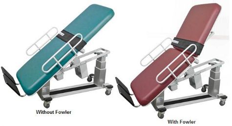
Enhancements to the table can reduce sonographer fatigue and require less repositioning of the patient, making it easy to get quality images in less time. An adjustable vascular scanning arm board can give easier access to the thyroid and the carotid arteries. Body straps can stabilize the patient and ensure safety throughout the entire procedure. Having an enhanced foot support can allow easier access to the patient?s calves without the need of the sonographer to crouch.
What Procedures can be Performed on an Ultrasound Table?
Lower extremity exams that can be performed on an ultrasound table include venous mapping and Doppler (DVT), venous reflux, venous insufficiency, venous ablation, and arterial scan/Doppler (PAD).
Upper extremity exams include arterial, venous access placement and assessment, carotid, carotid duplex, transcranial, abdomen, aorta, thyroid, breast, renal, renal transplant, renal artery, testicular, musculoskeletal, and superior mesenteric artery.
Mike Price, OT
Rehabmart Co-Founder & CTO
lb